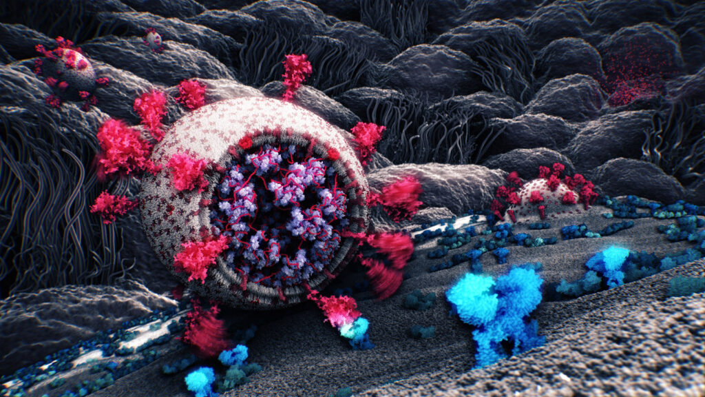
Making of a visualization of SARS-CoV-2
Tatjana Hirschmugl, Nanographics GmbH
Tobias Klein, Nanographics GmbH
Ondřej Strnad, KAUST
Deng Luo, KAUST
Peter Mindek, Nanographics GmbH, TU Wien
This image shows a part of the SARS-CoV-2 life-cycle, which happens in the epithelial cells of lungs of COVID-19 patients. We can see a virion’s spike protein binding to a receptor on the cell surface, another virion entering the cell by fusing with its membrane, and in the background, many new virions exiting an infected cell.